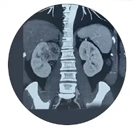
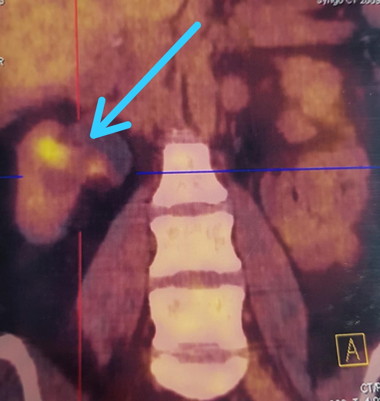
42 year old Omanian Female had an episode of blood in urine due to Kidney Cancer
Pellentesque porta sed dolor id volutpat. Vestibulum dolor turpis, consectetur at mauris eget.
Call/WhatsApp : +919136561220
Once patients present with symptoms as described above, a PLAIN CT SCAN abdomen or when a CT scan is unavailable, an Ultrasonography abdomen confirms presence of stone and absence of other pathology
We inject a radio opaque dye or contrast into the kidney. This visualizes each and every calyces (chambers) of the kidney under the C-arm. The endoscope is passed to each of the calyces to check if any residual stone is left anywhere. At the end of the procedure there should be no fragments more than 2mm. When we have confirmed this the kidney specialist puts in a stent and the procedure is over.
These methods involves perctaneous access/ placement of a tube from the back then through the abdominal wall into the kidney, to visualize the stone and then the stones are fragmented and dusted with laser and cleared.
The pelvicalyceal system/interior/lumen of kidney is visualized through an flexible ureterosrenoscopy, then the stones which are identified are fragmented and dusted with laser and cleared. The goal of holmium lithotripsy is to reduce stones to small fragments 2 mm or less. If the stone is large , the kidney is filled with dust and debris making visualization of fragments difficult, in such a situation a DJ Stent is placed and the stone is managed in a staged fashion in a second sitting after 2 weeks.
Can be done for stones less than 1cm , when they are in a favorable location in kidney ( non Lower pole) , and HU less than 1000
Rarely Laparoscopic assisted PNL may be required especially in the setting of PU junction obstruction.
Consult Dr Soumyan Dey today for Kidney Stone treatment. We are based in Mumbai, Navi Mumbai and Thane.
Pellentesque porta sed dolor id volutpat. Vestibulum dolor turpis, consectetur at mauris eget.